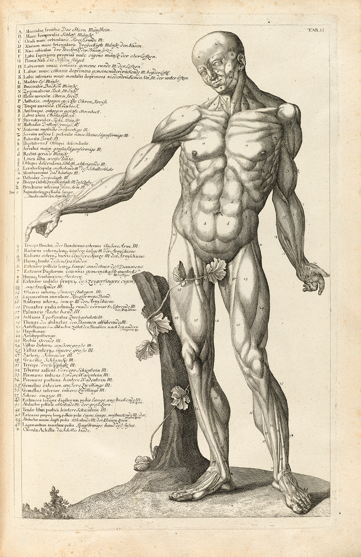
Body Muscles Labeled Front And Back : Muscle Anatomy Quiz : Fascia is the organ of structure.. 12 photos of the muscles labeled front and back. These lines wrap around muscles, separate muscle fibers, and muscles from bones, as well as connect them. The sartorius l the sartorius runs from the outside of the hip, down and across to the. This entry was posted in anatomy, muscles and tagged body, human muscle diagram, human muscles, muscle, muscles, muscles anatomy, muscles diagram, muscular system, s by admin. The coracobrachialis and pectoralis major muscles connect the humerus anteriorly to the scapula and ribs, flexing and adducting the arm toward the front of the body when you reach forward to grab an object.
Claim your free copy of the client back care guide today. New users enjoy 60% off. Together with several other muscles, the gluteus maximus muscles form the buttocks. This is a table of skeletal muscles of the human anatomy. There are 12 fascial lines in our body.
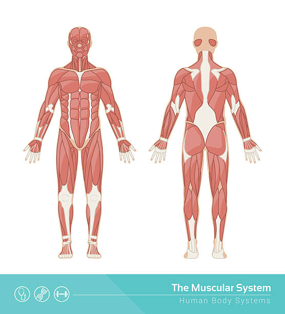
Historical Anatomies On The Web Browse Titles from www.nlm.nih.gov Link to client back care guide This entry was posted in anatomy, muscles and tagged body, human muscle diagram, human muscles, muscle, muscles, muscles anatomy, muscles diagram, muscular system, s by admin. See human back anatomy stock video clips. Use the location, shape and surrounding structures to help. Nine muscles of the chest and upper back are used to move the humerus (upper arm bone). Human body anatomy, front, back, side view, vector woman and man illustration, body silhouette. There are anterior muscles diagrams and posterior muscles diagrams. Posterior full body muscular system diagram.
These four muscles at the front of the thigh are the major extensors (help to extend the leg.
Posterior full body muscular system diagram. Triceps, biceps, pectoralis major, quadriceps, hamstrings, gluteus maximus, abdominals, deltoid, latissimus dorsi, external obliques, gastrocnemius, tibialis anterior. Nine muscles of the chest and upper back are used to move the humerus (upper arm bone). These four muscles at the front of the thigh are the major extensors (help to extend the leg. Quadriceps (made of 4 muscles): Human body anatomy, front, back, side view, vector woman and man illustration, body silhouette. The back supports the weight of the body, allowing for flexible movement while protecting vital organs and nerve structures. Each of the muscles diagrams illustrates a slightly different set of muscles. Claim your free copy of the client back care guide today. It is the overall fabric that makes up our body. There are 12 fascial lines in our body. Link to client back care guide Brings shoulders and arms back to body.
Unlabeled muscular system front and back. Labeled illustration chart on white. Download 178 man body anatomy front back side stock illustrations, vectors & clipart for free or amazingly low rates! Posterior full body muscular system diagram. New users enjoy 60% off.
List Of Human Anatomical Regions Wikipedia from upload.wikimedia.org Lower thoracic, lumbar vertebrae and sacrum: Here my aim is simply to bring these major lines to your attention. Related posts of muscle anatomy front and back muscle anatomy stomach. Front of human upper leg: The gluteus maximus is the largest muscle in the body. It is controlled by the axillary nerve. Anatomy of the nose and throat. Fascia is the organ of structure.
This is a table of skeletal muscles of the human anatomy.
It allows for movement of the shoulders and shoulder blades.distal phalanx of the big toe. The coracobrachialis and pectoralis major muscles connect the humerus anteriorly to the scapula and ribs, flexing and adducting the arm toward the front of the body when you reach forward to grab an object. Posterior full body muscular system diagram. Claim your free copy of the client back care guide today. Your clients will thank you for it! The gluteus maximus is the largest muscle in the body. Moves humerus (arm) to chest. Human body anatomy female female anatomy muscle shoulder blade pain anatomy back muscles bones man female anatomy body muscles in a body female anatomy muscole shoulder concept muscular sysyem. New users enjoy 60% off. The muscles that make up the quadriceps are the strongest and leanest of all muscles in the body. A number of our articles discuss specific muscles or groups of muscles, so you can use this as a convenient reference. By tightening and relaxing, the skeletal muscles create movement. Brings shoulders and arms back to body.
Together with several other muscles, the gluteus maximus muscles form the buttocks. You use this muscle when you stand up, walk, run, and climb stairs in fact—whenever you straighten or extend your legs. The sartorius l the sartorius runs from the outside of the hip, down and across to the. Labeled illustration chart on white. Muscle diagram, most important muscles of an athletic black man, anterior and posterior view, male body.
219 780 Human Muscle Stock Photos Pictures Royalty Free Images Istock from media.istockphoto.com This is a table of skeletal muscles of the human anatomy. These lines wrap around muscles, separate muscle fibers, and muscles from bones, as well as connect them. Here my aim is simply to bring these major lines to your attention. By tightening and relaxing, the skeletal muscles create movement. They support bones, in this case, the vertebrae. Use the location, shape and surrounding structures to help. Human body anatomy, body silhouette. The back supports the weight of the body, allowing for flexible movement while protecting vital organs and nerve structures.
Related posts of muscle anatomy front and back muscle anatomy stomach.
Moves humerus (arm) to chest. Unlabeled muscular system front and back. Female cardiovascular system, rear and front views, on black. Your clients will thank you for it! Front of human upper leg: In this image, you will find frontalis, orbicularis oculi, zygomaticus, masseter, orbicularis oris, sternocleidomasteoid, deltoid, pectoralis major, biceps brachii, iliopsoas, adductor longus, gastrocnemius. On the posterior side of the arm the teres major. They support bones, in this case, the vertebrae. Related posts of muscle anatomy front and back muscle anatomy stomach. Human body anatomy, body silhouette. It runs from the back of the pelvis to the upper part of the femur. The muscles of the lower back, including the erector spinae and quadratus lumborum muscles, contract to extend and laterally bend the vertebral column. 12 photos of the muscles labeled front and back.


0 Comments